A. Cerebri Media Anatomy | The shape of the gland resembles a pine cone, which gives it its name. The mca supplies many deep brain structures, the majority of the lateral surface of the cerebral hemispheres, and the temporal pole of the brain. Each primary motor cortex sends instructions for. The brain is one of the most highly perfused organs in the body. Another, the tentorium cerebelli, provides a strong, membranous roof over the cerebellum
The precentral gyrus contains the primary motor cortex (brodmann's area 4), which is responsible for integrating signals from different brain regions to modulate motor function: The pineal gland, conarium, or epiphysis cerebri, is a small endocrine gland in the brain of most vertebrates. It is therefore not surprising that the arterial blood supply to the human brain consists of two pairs of large arteries, the right and left internal carotid and the right and left vertebral arteries (figure 1). The dura mater is partitioned into several septa, which support the brain. Another, the tentorium cerebelli, provides a strong, membranous roof over the cerebellum
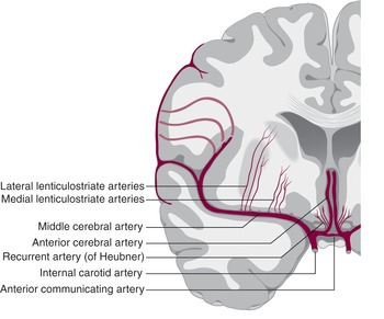
The dura mater is partitioned into several septa, which support the brain. May 31, 2021 · the precentral gyrus is situated between and parallel to the central and precentral sulci, and is the most posterior structure considered part of the frontal lobe. May 31, 2021 · middle cerebral artery (arteria cerebri media) the middle cerebral artery (mca) is a terminal branch of the internal carotid artery and is part of the anterior cerebral circulation. The internal carotid arteries principally supply the cerebrum, whereas the two vertebral arteries join distally to form. Although the text went on to have tremendous impact on neurological sciences and anatomy, willis initially published the text pursuant to his understanding of the philosophical soul. The precentral gyrus contains the primary motor cortex (brodmann's area 4), which is responsible for integrating signals from different brain regions to modulate motor function: Each primary motor cortex sends instructions for. Aug 15, 2017 · the "circle" was first described in a book written by dr. The pineal gland, conarium, or epiphysis cerebri, is a small endocrine gland in the brain of most vertebrates. The brain is one of the most highly perfused organs in the body. Jun 30, 2016 · the ventricles of the brain are a communicating network of cavities filled with cerebrospinal fluid (csf) and located within the brain parenchyma. The mca supplies many deep brain structures, the majority of the lateral surface of the cerebral hemispheres, and the temporal pole of the brain. A basic understanding of skin anatomy is important when explaining the process of skin biopsy.
The mca supplies many deep brain structures, the majority of the lateral surface of the cerebral hemispheres, and the temporal pole of the brain. Aug 15, 2017 · the "circle" was first described in a book written by dr. A basic understanding of skin anatomy is important when explaining the process of skin biopsy. Another, the tentorium cerebelli, provides a strong, membranous roof over the cerebellum Jun 30, 2016 · the ventricles of the brain are a communicating network of cavities filled with cerebrospinal fluid (csf) and located within the brain parenchyma.

May 31, 2021 · middle cerebral artery (arteria cerebri media) the middle cerebral artery (mca) is a terminal branch of the internal carotid artery and is part of the anterior cerebral circulation. Each primary motor cortex sends instructions for. The ventricular system is composed of 2 lateral ventricles, the third ventricle, the cerebral aqueduct, and the fourth ventricle (see the following images). The internal carotid arteries principally supply the cerebrum, whereas the two vertebral arteries join distally to form. Thomas willis in 1664, cerebri anatome. Jun 30, 2016 · the ventricles of the brain are a communicating network of cavities filled with cerebrospinal fluid (csf) and located within the brain parenchyma. The brain is one of the most highly perfused organs in the body. May 31, 2021 · the precentral gyrus is situated between and parallel to the central and precentral sulci, and is the most posterior structure considered part of the frontal lobe. The mca supplies many deep brain structures, the majority of the lateral surface of the cerebral hemispheres, and the temporal pole of the brain. Another, the tentorium cerebelli, provides a strong, membranous roof over the cerebellum A basic understanding of skin anatomy is important when explaining the process of skin biopsy. The shape of the gland resembles a pine cone, which gives it its name. The falx cerebri attaches anteriorly at the crista galli in proximity to the cribriform plate and to the frontal and.
The mca supplies many deep brain structures, the majority of the lateral surface of the cerebral hemispheres, and the temporal pole of the brain. Jun 30, 2016 · the ventricles of the brain are a communicating network of cavities filled with cerebrospinal fluid (csf) and located within the brain parenchyma. The precentral gyrus contains the primary motor cortex (brodmann's area 4), which is responsible for integrating signals from different brain regions to modulate motor function: The internal carotid arteries principally supply the cerebrum, whereas the two vertebral arteries join distally to form. The shape of the gland resembles a pine cone, which gives it its name.

Although the text went on to have tremendous impact on neurological sciences and anatomy, willis initially published the text pursuant to his understanding of the philosophical soul. The falx cerebri attaches anteriorly at the crista galli in proximity to the cribriform plate and to the frontal and. The shape of the gland resembles a pine cone, which gives it its name. Each component of the skin plays a role in its daily function, therefore every component is a source of vital information that can be captured and assessed with a skin biopsy. The brain is one of the most highly perfused organs in the body. Each primary motor cortex sends instructions for. It is therefore not surprising that the arterial blood supply to the human brain consists of two pairs of large arteries, the right and left internal carotid and the right and left vertebral arteries (figure 1). The pineal gland, conarium, or epiphysis cerebri, is a small endocrine gland in the brain of most vertebrates. Thomas willis in 1664, cerebri anatome. Jun 30, 2016 · the ventricles of the brain are a communicating network of cavities filled with cerebrospinal fluid (csf) and located within the brain parenchyma. The ventricular system is composed of 2 lateral ventricles, the third ventricle, the cerebral aqueduct, and the fourth ventricle (see the following images). The mca supplies many deep brain structures, the majority of the lateral surface of the cerebral hemispheres, and the temporal pole of the brain. The precentral gyrus contains the primary motor cortex (brodmann's area 4), which is responsible for integrating signals from different brain regions to modulate motor function:
A basic understanding of skin anatomy is important when explaining the process of skin biopsy a. cerebri media. It is therefore not surprising that the arterial blood supply to the human brain consists of two pairs of large arteries, the right and left internal carotid and the right and left vertebral arteries (figure 1).
A. Cerebri Media Anatomy! The falx cerebri attaches anteriorly at the crista galli in proximity to the cribriform plate and to the frontal and.